
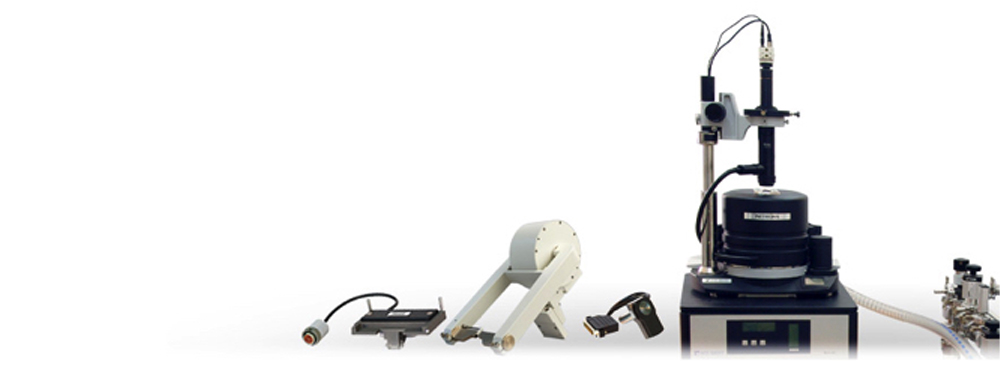
NTEGRA MFM
AFM for studies with in-plane and out-plane external magnetic fields
The principle of Magnetic Force Microscopy (MFM) is based on the detection of the interaction between the sample and a nanosized magnetic probe. The standard magnetic probeis an AFM cantilever covered by thin magnetic film. MFM measurements reveal magnetic structure ofthin films, bulk samples, nanostructures and nanoparticles with resolution down to nanometer scale. The best resolution is achieved by using special high-aspect ratio tips. There are two main methods of MFM signal detection: measurements of static cantilever deflection and dynamic MFM detecting amplitude, phase and frequency of oscillating cantilever. Standard MFM methods are available with all SPM models produced by NT-MDT
Due to the open architecture, the functionality of NTEGRA Aura can be widen essentially: specialized magnetic measurements with external magnetic field (horizontal, up to +/-0.3 T; vertical, up to +/-0.08 T), high-temperature experiments (heating up to 150 °С with temperature maintaining precision of 0.05 °С), etc.
APPLICATION EXAMPLES
The use of an in-situ magnetic field during MFM measurements allows the investigation of magnetization reversal processes.
Horizontal external magnetic field
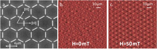
MFM images of artificial spin ice (honeycomb magnetic structure shown in image (a)): (b) demagnetized state, (c) the same place with highly ordered structure in horizontal magnetic field of 50 mT applied in the [11] direction. Schumann, et al. Appl. Phys. Lett. 97, 022509 ,2010
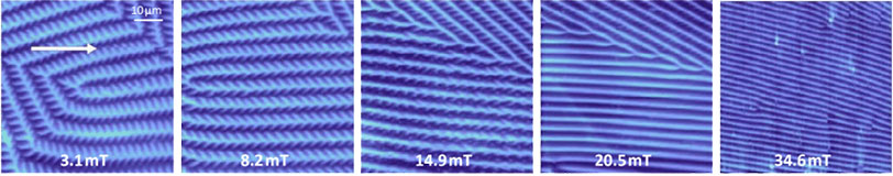
Reorganization of the garnet film domain structure in external horizontal magnetic field. Direction of field is indicated by arrow.
Vertical external magnetic field
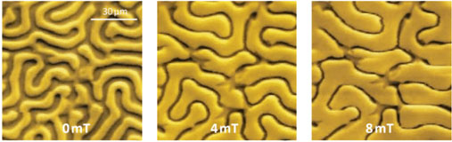
Figure 7. MFM images of garnet film in vertical magnetic field. Extension of domains with magnetization direction coinciding with direction of the external field is clearly seen. Sample courtesy: Prof. F.V. Lisovsky (IRE RAS, Russia)
SPECIFICATIONS
- AFM (contact + intermittent contact)
- HybriD mode
- Lateral Force Microscopy
- Force Modulation Microscopy
- Magnetic Force Microscopy
- Electrostatic Force Microscopy
- Scanning Capacitance Force Microscopy
- Kelvin Probe Force Microscopy
- Piezoresponce Force Microscopy
- Spreading-Resistance Imaging
- STM
- Nanosclerometry
- Lithography: AFM (Force + Current), STM
TECHNICAL DATA
| External magnetic field | Horizontal up to +/-0.3 T; vertical, up to +/-0.08 T |
|
| Scan type | Scanning by probe* | |
| Sample size | Up to 100 mm in diameter, up to 15 mm in height |
|
| Sample weight | Up to 300 g | |
| XY sample positiniong range | 5×5 mm | |
| Positioning resolution | readable resolution – 5 um sensitivity – 2 um |
|
| Scan range | 100x100x10 um | |
| Scan range | Up to 200x200x15 um (DualScan mode) | |
| Non linearity, XY (with closed loop sensors) |
≤0.15% | |
| Noise level, Z (RMS in bandwidth 1000 Hz) |
With sensors | 0.06 nm (typically), ≤0.07 nm |
| Noise level, Z (RMS in bandwidth 1000 Hz) |
Without sensors | 0.05 nm |
| Noise level, XY** (RMS in bandwidth 200 Hz) |
With sensors | 0.1 nm (typically), ≤0.2 nm |
| Noise level, XY** (RMS in bandwidth 200 Hz) |
Without sensors | 0.01 nm |
| Optical viewing system | Optical resolution | 3 um |
| Optical viewing system | Field of view | 2.0-0.4 mm |
| Optical viewing system | Continuous zoom | available |
| Temperature control | Range | From RT to +150 °C |
| Temperature control | Stability | ±0.005 °C (typically), ≤±0.01 °C |
| Vibration isolation | Active | 0.7-1000 Hz |
| Vibration isolation | Passive | above 1 kHz |
* Scanning head can be configured to serve as a stand-alone device for specimens of unlimited sizes.
** Built-in capacitive sensors have extremely low noise and any area down to 50×50 nm can be scanned with closed-loop control.